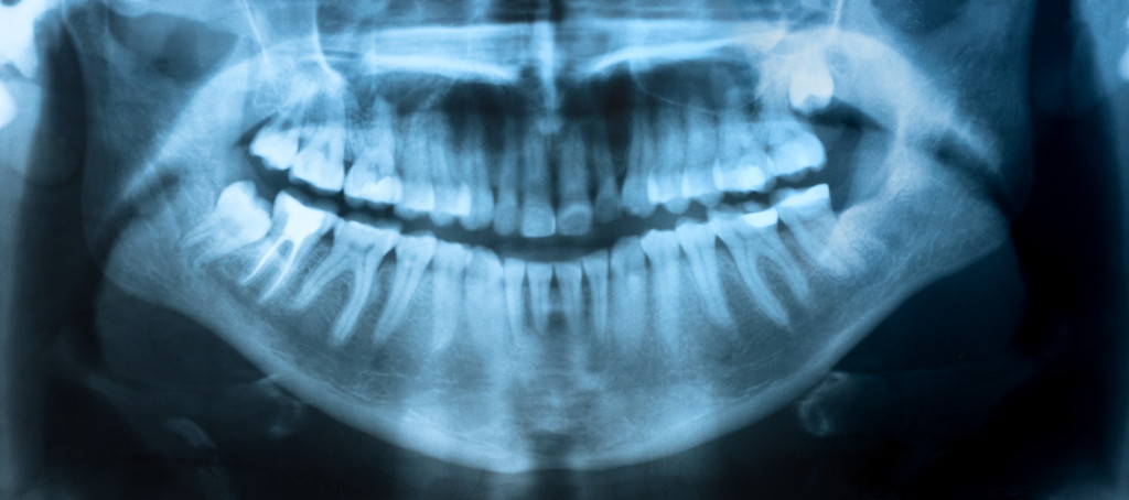
Digital radiography (often referred to as digital X-ray) is a radiography technique that uses electronic sensor instead of photographic film. Thanks to this, digital X-ray has two important advantages over traditional X-ray imaging. Firstly, unlike with traditional X-ray technology, the image is available almost instantly, which makes the use of this diagnostic method much more convenient. Secondly, the created digital image is much more precise and detailed in comparison to traditional photographic film. This allows dentists to spot certain changes that would have been missed by looking at traditional X-ray images. Also, digital image can be stored on computer and it can be transferred much quicker and easier if needed. The imaging principle is the same as with traditional X-ray imaging technique: the X-ray pass through the body and are registered by the sensor. More dense structures, such as healthy teeth, allow less radiation to pass through; less radiation is detected by the sensor, which is why these structures appear lighter on the image. Less dense structures, such as dental caries, allow more radiation to go through and be registered by the sensor, which makes them appear darker. That is how dentists interpret X-ray images.
Just as traditional X-ray imaging, digital X-ray exposes the patient to very low radiation doses, which makes this diagnostic method very safe. Nevertheless, additional protection may be used in some situations. The protection comprises leaded aprons and thyroid collars, and is usually used as an additional precautionary measure for children and women of childbearing age. Nevertheless, digital X-ray imaging is in any case one of the safest diagnostic methods in dentistry.
You can call us at 718-255-8555 Mon – Fri 9am – 7pm, email us at support@dentalxo.com or fill out following appointment request form HERE